


CASE PRESENTATION
Elevating Diagnostics with PSP Technology
Back in 2006, I was so disappointed and uncomfortable with the images I was receiving from other dental offices. I simply could not see the anatomy necessary for a proper diagnosis, so I set out to invest in a system that was far superior. The ScanX Swift digital radiography system from Air Techniques has been the answer. Compared to thick, inflexible hardwired sensors, patients find phosphor storage plates (PSPs) to be much more comfortable. But what is even more impactful is how much surface area is exposed on each individual PSP image—meaning fewer images and less radiation are needed.
Here are just a few case examples that show how ScanX PSP technology has given me the opportunity to do more dentistry while impressing patients on their first visits.
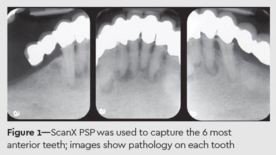
Case 1—Imaging without Limits
A patient wearing an extensive roundhouse bridgework presented to our office in pain. I easily captured closeup images of the 6 most anterior teeth, which showed pathology on each tooth (Figure 1). Realizing that 6 root canals were needed, I referred the patient to an endodontist so that treatment could be expedited.
These ended up being very difficult root canals, so I was glad to have referred them out. The endodontist did a beautiful job, but with a catch—the radiography was completely lacking. When the patient returned to my office for his restorations, only 5 teeth appeared on the x-ray out of 6 root canals that were completed, and only 2 apices were evident (Figure 2). There was also a big area of dark, black space at the top of the x-ray because the patient had a small mouth and the hardwired sensor could not fit all the way down.

I performed 6 post and cores, recemented the roundhouse bridge, and sent the completed radiograph to the insurance company. The response I received back was a dreaded one: “We’re sorry, you have not adequately demonstrated that root canal was properly completed on all 6 teeth on this patient.”
Using a ScanX PSP, I captured one x-ray that was not elongated and showed all 6 root canals on the individual image (Figure 3). I then sent the image to both the insurance company and the endodontist for his records. I received a check from the insurance company and a question from the endodontist: “How did you get all the teeth in one shot?”

Case 2—Seeing Beyond the Apex
A patient presented with some pain in his mandibular right quadrant. While percussing the teeth, I found that tooth No. 29, the second premolar, had pulpitis, and that the pain he was experiencing was from a very large and deep amalgam. He had huge mandibular tori that may have presented a challenge using traditional hardwired sensors, but we had no issue fitting a soft, flexible ScanX PSP sensor in his mouth. The tori were directly visible in the radiograph and you could see not only the apex of the tooth, but almost a centimeter beyond the tip of the root. Because these images were so easily captured without the need for a panoramic x-ray, the root canal commenced easily (Figures 4-6).


Case 3—A Genius Diagnosis
A patient presented with significant pain in her upper left quadrant. At her previous dentist, she had 2 x-rays taken with a hardwired sensor where you could not see the distal buccal root of her tooth No. 15, 3-rooted second molar. Even though it was not a complete x-ray, the dentist advised that she may need a root canal on tooth No. 15, which made her feel uncomfortable.
Because ScanX PSPs are so easy to place, I captured not only tooth No. 15, but the space behind it (Figure 7), which revealed that there was nothing wrong with the second molar and that the problem was with the patient’s third molar. The tooth had a tiny conical root, and I was able to extract it in 2 minutes. So, the patient didn’t need a root canal after all—the problem was as simple as popping out a little wisdom tooth.

The patient was so impressed that I found the problem that it made me appear to her that I was some sort of a diagnostic genius, when all I did was expose a proper x-ray. She has been my loyal patient now for nearly 5 years. With the incredible ease of using ScanX PSPs, the diagnostics appear to patients as brilliant and beyond the norm when, in fact, they’re incredibly easy to take and keep patients comfortable.
James G. Kouzoukian, DDS
Dr. James G. Kouzoukian is a 1984 graduate of the New York University College of Dentistry. He has over 30 years of experience working with attorneys as an expert witness in many types of dental-legal matters. Dr. Kouzoukian is an internationally recognized lecturer and has presented numerous nationwide live seminars and several worldwide webinars on topics including TMJ dysfunction, 2D and 3D digital radiographic imaging, dental professional liability, oral hygiene, and electronic submission of third-party dental claims. He is president of the Institute for Continuing Dental Education and an active member of the board of trustees at the Queens County Dental Society, a division of the New York State Dental Association.
 Go-To Product Used in this Case
The ScanX Swift digital radiography system delivers crisp, sharp images in up to 20 lp/mm true resolution to meet the needs of all diagnostic requirements. ScanX PSPs have a 100% active surface area, which means users can capture up to nearly 40% more anatomy than with a comparable sized wired sensor.
