



By Lou Shuman, DMD, CAGS
HIGHER-QUALITY RESTORATIONS
Impression accuracy is directly connected to accurate restoration fit. Distortions, voids or a bad fit of traditional impressions can compromise periodontal health, function and longevity of the restoration, which means more frustrating redos and appointments. As noted by Dhull and colleagues, intraoral scanners capture 3D images of the mouth using sophisticated optics, imaging sensors and software algorithms.
Intraoral scanners aren’t without their complications. Alkadi describes the accuracy of intraoral scans as being influenced by factors that include operator skill and the patient’s anatomy. For these reasons, scanner selection is one of the most important factors for eliminating much of the margin for error.
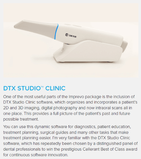
DEXIS recently updated its wireless scanner, the DEXIS Imprevo. The DEXIS 3800 series already offered a light, fast and simple-to-use scanner. However, the Imprevo scanner tip is 20% slimmer than its predecessor and it’s autoclavable, offering precision, speed and ease of disinfection. The DEXIS team, which designed the Imprevo upgrade, has a global reputation for imaging excellence.
Optics serves as one of the Imprevo’s most impressive aspects. With a 25-mm depth of field (39% greater than the previous generation), the scanner can access deeper impressions and tight interproximal spaces. Its “dual sensor technology” combines a laser, which produces a better and brighter light source, with white LED lighting for sharper and ultra-detailed images.
The improved capture of realistic colors and textures lets clinicians see hard and soft tissues better. The scanner features “enhanced texture capture,” which integrates mesh and texture, improving margin definition. Having 3D images plays an important role in patient communication. Clinicians not only benefit from the diagnostic clarity, but they can merge high-resolution textures onto the 3D mesh and show the realistic digital renderings to patients, which can aid communication that leads to treatment acceptance.
Within the DTX Studio™ Clinic platform (included with the Imprevo), AI in the SmartFusion feature can automatically merge intraoral texture/mesh scans with CBCT data to provide a comprehensive 3D view of the patient’s underlying anatomy.
IT’S A GOOD FIT
Dentistry continues to evolve, making it difficult to decide which innovations to add to the treatment experience. Enhancing patient comfort, improving the restorative process and capturing more accurate data for a better prosthetic design should be a clear “yes” from solo to multi-practitioner practices. Digital scanning with Imprevo creates a literal “good fit” and will certainly make a good impression on the practice and the patients.
REFERENCES
Alkadi L. A comprehensive review of factors that influence the accuracy of intraoral scanners. Diagnostics. 2023;13(21):3291. doi:10.3390/diagnostics13213291
Versaci MB. ACE Panel report finds about half of dentists use intraoral scanners. ANA News. September 21, 2021. adanews.ada.org/ada-news/2021/september/ace-panel-report-finds-about-half-of-dentists-use-intraoral-scanners
Dhull KS, Nagar R, Mathur P, et al. Intraoral scanners: Mechanism, applications, advantages, and limitations. J Pharm Bioallied Sci. 2024;16(Suppl 3):S1929-31. doi:10.4103/jpbs.jpbs_1299_23