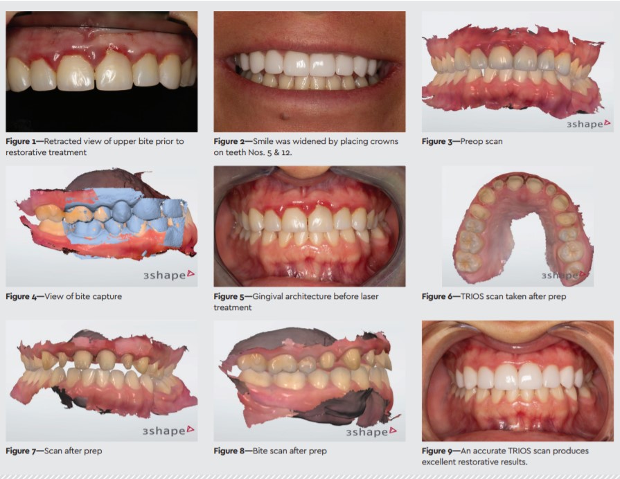
Case Presentation: Predictable Restorations with 3Shape TRIOS
Since I adopted the 3Shape TRIOS intraoral scanner, it has been a win-win for my patients and my practice. Compared to traditional analog impressions, digital impressions have a plethora of benefits that include predictable results, improved patient comfort, accurate scans, quicker appointments, quicker lab turnaround time, and better-fitting final restorations that usually require no adjustments. As a result, the practice’s ROI has improved dramatically, making the TRIOS scanner one of the best investments I have made.
A 27-year-old female patient reported that she was unhappy with the fi t and appearance of her existing zirconia crowns and with the color of her teeth. Intraoral x-rays (KaVo FOCUS, KaVo Imaging) revealed 6 ill-fitting crowns on teeth Nos. 6–11 with open margins on Nos. 7–10 (Figure 1). After discussing treatment options with the patient, we decided to remove and replace all 6 crowns and place 2 new crowns on teeth Nos. 5 and 12 to widen her smile (Figure 2).
Efficiency, Convenience, Accuracy
The patient was photographed, and the upper, lower, and bite arches were scanned in full color (Figures 3 & 4) with the TRIOS intraoral scanner (3Shape). The cordless design, compact size, and smart tip of the scanner ensure that scans are taken quickly, efficiently, and accurately. Unlike traditional impressions, TRIOS scans minimize the possibility of distortion during the impression-taking process and are comfortable for patients. After administering a local anesthetic (Septocaine, Septodont), a diode laser (Biolase) was used to reshape the gingival architecture (Figure 5).
Before beginning the restorative process, I took alginate impressions (Primo, TriState Dental) of the upper arch, which were used to create the provisional restorations. I removed the 6 old crowns with a coarse grit diamond bur (NTI Diamonds, Kerr) and redefined the preps using a series of friction-grip diamond burs (Brasseler). Bleeding was controlled with ViscoStat Clear (Ultradent).
Scan for Bite Capture Details
After redefining the preparations and prepping for 2 additional crowns, we again used the TRIOS to digitally scan the preparations (Figure 6). The 3Shape software automatically captured the bite from the initial scans (Figures 7 & 8). After the final scan, we used Primo Temporary Crown and Bridge Material (TriState Dental) with the previously taken alginate impressions to create the initial phase of the temporary provisionals. From there we redesigned, reshaped, and corrected the restorations using Tetric EvoFlow flowable composite and IPS Empress Direct (Ivoclar Vivadent) and a series of carbide burs to give us proper occlusion and esthetics. We cemented the temporary provisionals with TempoCem (DMG America). The patient was sent home with plans to return in 2 to 3 weeks for the final steps in the restorative process.
Instantaneous Digital Transmission
Using the 3Shape software, digital transmission to the laboratory is instantaneous, and the intraoralscans and photographs were quickly transmitted to the lab (Precision Dental Products; Draper, UT). We received the 8 finished ceramic crowns from the lab within 2 to 3 weeks.
A Perfect Fit
The patient returned for removal of the temps, and 8 IPS e.max ceramic crowns (Ivoclar Vivadent) were successfully placed. Because of the accuracy of the TRIOS scans, few to no adjustments were needed, and the crowns fit perfectly. The final results revealed a beautiful smile (Figures 2 & 9), and the patient was delighted with her smile makeover.