A 26-year-old man was referred to our office for evaluation of the maxillary left first molar, tooth No. 14, which presented with advanced periodontal involvement.

Dr. Peterson is a board-certified periodontist and periodontal surgeon at Arcadia Perio in Arcadia, CA. He earned his dental degree and specialty training in periodontics from Oregon Health & Science University. After beginning his career in private practice in the Portland area, he served as an adjunct professor in the Graduate Periodontics program at Loma Linda University before returning to full-time clinical practice. Dr. Peterson has extensive experience in periodontal regeneration and implant dentistry. He is a certified LANAP clinician and instructor through the Institute for Advanced Laser Dentistry, and he frequently lectures on advanced periodontal and implant techniques. His clinical interests include digital implant workflows and X-Nav guided implant placement.
A 26-year-old man was referred to our office for evaluation of the maxillary left first molar, tooth No. 14, which presented with advanced periodontal involvement.
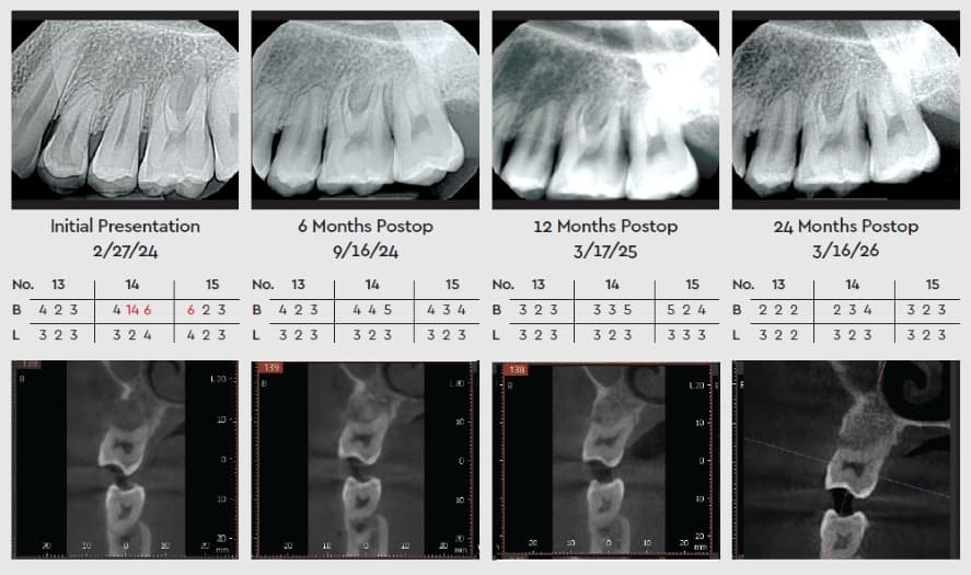
Clinical examination revealed Grade II buccal furcation involvement with buccal pocket depths of 4–14–6 mm and lingual depths of 3–2–4 mm, along with radiographic evidence of bone loss extending into the furcation.
Given the patient’s young age and the extent of periodontal destruction localized to this tooth, the long-term prognosis was considered guarded and heavily dependent on our ability to halt and control disease progression. The referring dentist had discussed the possibility that No. 14 might ultimately require extraction if the periodontal condition could not be stabilized.
The Plan: Maximize True Regeneration
After periodontal evaluation and risk assessment, we elected to treat the patient with the LANAP (Laser Assisted New Attachment Procedure) protocol, which is only available using a specialized Nd:YAG dental laser (PerioLase MVP-7, Millennium Dental Technologies).
The LANAP protocol’s ability to selectively remove diseased epithelium and infected pocket lining—while disinfecting the pocket and promoting a stable, fibrin-rich clot—made it well-suited for a furcation defect in a young patient where long-term tooth preservation was a priority. This laser-based approach was chosen over traditional flap surgery to maximize potential for true regeneration, minimize surgical trauma, and preserve as much natural tooth structure and supporting tissue as possible.
The LANAP Procedure
LANAP therapy was performed with standard protocol, including initial laser decontamination of the pocket, thorough ultrasonic root debridement, and a second laser pass to stabilize the wound and encourage new attachment. No scalpel incisions or sutures were required, and the patient reported minimal post-op discomfort. Occlusal forces were evaluated and adjusted as needed to decrease mechanical stress on the compromised molar, further supporting periodontal healing.
The case was carefully documented and followed over time. At 6 months, clinical re-evaluation showed a marked reduction in inflammation, decreased probing depths, and improved tissue tone in the furcation area, with 2D radiographs already suggesting early bone fill within the buccal furcation.
Periodontal Healing Over Time
At 12 months, pocket depths around No. 14 had normalized into a maintainable range, bleeding on probing was absent, and the tooth demonstrated improved stability. Radiographically, there was clear evidence of increased radiopacity in the furcation region consistent with bone regeneration.
At 24 months, 2D radiographs and 3D imaging confirmed substantial bone regeneration into the previously compromised furcation. The defect that had once been a clear Grade II buccal furcation involvement now demonstrated significant radiographic fill and a much more favorable architecture.
Clinically, No. 14 was stable, functional, and asymptomatic, with probing depths in a healthy, maintainable range and no signs of recurrent inflammation. The tooth’s prognosis—originally considered guarded and questionable for long-term retention—was greatly improved and is now classified as favorable, contingent on continued periodontal maintenance and home care.
The Power of PerioLase
This case highlights the effectiveness of the LANAP protocol in managing challenging periodontal defects, especially furcation involvement in a young patient where extraction and replacement would have been a significant lifelong burden.
The LANAP protocol reduces harmful bacteria while preserving healthy tissue. By promoting immediate blood clot formation, it acts as a natural bandage to reduce pain, bleeding, and swelling—leading to a faster and more comfortable recovery compared with traditional osseous surgery.
Ultimately, LANAP therapy using the PerioLase MVP-7 laser allowed a minimally invasive, regenerative approach that not only preserved the tooth but also demonstrated true radiographic and clinical improvement over time.
For referring providers, this case exemplifies how modern laser periodontal therapy can convert a borderline tooth into a stable, long-term asset, supported by objective documentation with both 2D and 3D imaging.