Dr. Springmann
has practiced
general dentistry in
Williamsburg, VA, for
25 years. She trained
as a biomedical
sculptor at New
York University
and received her
Doctor of Dental
Surgery Degree
from the Medical
College of Virginia.
Dr. Springmann
has earned her
Fellowship in the
Academy of General
Dentistry.
Every dental professional seeks to provide patients with the best long-term outcomes, both esthetically and functionally. My practice strives to incorporate the latest technology so I can best communicate with my patients and laboratory technicians. Using a digital dental camera has provided me the ability to obtain high-quality digital images so I can better execute cosmetic cases, document pathology, and monitor changes in the oral cavity, among many other applications.
Case Presentation
A female patient with an unremarkable health history presented to my practice with a chief complaint of poor dental esthetics. A full dental exam was performed and treatment options were discussed with the patient. The patient elected to have Invisalign treatment and existing crowns replaced on teeth Nos. 7 through 10.
Prior to initiation of Invisalign treatment, tooth No. 13 was fractured. After a discussion of treatment options, the patient elected for extraction and implant placement. The missing tooth can be seen in Figure 2. Space for implant placement was maintained with a pontic in the patient’s Invisalign aligners.
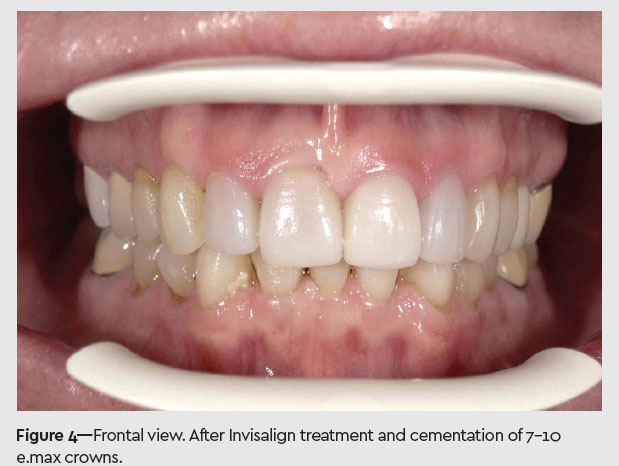
The patient’s Invisalign treatment lasted approximately 16 months and was followed by implant placement in site 13. All intraoral photo documentation was made with the EyeSpecial C-II digital camera (Shofu), with the exception of the patient’s pre-Invisalign photos due to the offi ce purchasing the camera during her treatment. The EyeSpecial C-II camera is used frequently in cosmetic cases to relay information about shade and esthetics to the lab. Figure 1 shows the patient’s previously existing crowns, and Figure 4 shows the newly fabricated e.max crowns. As with all my Invisalign cases, digital images were used to communicate patient’s occlusion, facial profi le, and esthetics (Figures 1 to 6).
Next, crowns 7 through 10 were removed and the teeth were prepared for IPS e.max crowns (Ivoclar Vivadent).
Whitening
Outside of this case, the EyeSpecial C-II is also used to document shade progress for whitening cases. I use Philips Zoom whitening because it is an optimal in-offi ce whitening system for patients who want to “jump-start” their whitening. Figures 7 and 8 show our clinical setup using Philips Zoom WhiteSpeed. Each treatment usually consists of four to fi ve 15-minute treatments. The whitening process is accelerated when the blue LED light-activated technology and whitening gel are used together. Often, I encourage use of a 5,000 ppm dentifrice before treatment to help manage sensitivity. Figures 9 and 10 show the same-day “before” and “after” of a different patient who had ZOOM treatment. Patients are provided with custom trays and
Philips Zoom take-home whitening gel so they can continue to whiten at their pace to suit their own needs.