


Protect Your Patients’ Oral & Overall Health with CBCT Imaging
It’s not every day that a patient says, “Dr. Clark, if you hadn’t taken that scan, I might not be here today.” Yet those were the exact words of a patient who presented for a consultation to address her TMD symptoms, which included frequent headaches, chronic ear congestion, vertigo, TMJ popping and crepitus, and chronic neck and shoulder pain. Her health was further impacted by several well-monitored medical conditions, including hyperlipidemia, hypertension, and type 2 diabetes, along with anxiety, mild obstructive sleep apnea, and gastroesophageal reflux disease.
She’d previously had TMD treatment from another doctor and was wearing an orthotic to help reposition her bite and alleviate some of her frequent symptoms, such as headaches and facial pain. She’d been in the orthotic for a while and it was starting to break down, which changed the position of her bite and caused her TMD symptoms to resurface.
A Life-Saving Discovery
To gain a comprehensive understanding of the complete disposition of a presenting TMD patient, it is important to image all the structures that may affect or be affected by the condition. Thus, a 26 × 30 cm Ultra Low Dose, 300-micron resolution volume was captured with our Planmeca Viso G7 CBCT imaging unit. This is our protocol for any patient who comes in with TMD symptoms, headaches, or sleep apnea issues.
On this patient’s scan, a roughly 95% atheromatous blockage at the bifurcation of the right carotid artery was noted and verified by oral radiologist Peter Green, DDS, Raleigh, NC, as any dental clinician’s finding that is questionable can be forwarded for review by a radiologist for a nominal fee. Another large atheroma was noted on the left side, but it did not completely encircle the artery and was of less concern. After discussion and education on the findings with the patient, she was referred to her cardiologist. A copy of the DICOM was given to the patient, accompanied with a viewer application to allow other providers to review the scan. All dental or TMD treatments were postponed until resolution and release from her team of physicians.
Successful Treatment Outcome
After consultation with the patient’s cardiologist, she was referred to a vascular surgeon for evaluation and planning. An endarterectomy was performed by Adam Richter, MD, FACS, RPVI, of The Surgical Clinic in Nashville, TN, to remove the lesion at the bifurcation of the right carotid artery. After release from her physicians, she returned to initiate dental treatment and TMD therapy.
We have since made her a new orthotic and her symptoms have subsided. She has a few missing teeth, so our goal is to fabricate a long-term device that will serve as a partial denture while repositioning her bite at the same time.
Every Patient Deserves a Planmeca Ultra Low Dose Scan
I take a large-volume CBCT scan with the Planmeca Viso G7 on every single patient who walks into the office—even if it’s a routine new patient—because the dose is similar to that of a pan and significantly less than a full-mouth series. And I get so much more important information that I can see in three dimensions.
The Power of CBCT
Because of how often I capture these large-volume scans with the Planmeca Viso G7, I see carotid atheromas in various areas around 5 to 10% of the time. But I almost never see them as big and with this much of an arterial blockage as I did in this case. This patient was completely surprised at the findings of this scan, as she was not actively experiencing symptoms of an artery blockage.
This just goes to show that there is a wealth of asymptomatic dental pathology out there that is truly mind-blowing, and unfortunately, we just can’t identify it on 2D radiographs alone. If you strive for a practice where you are invested in your patients’ overall health as well as comprehensive dentistry, then a CBCT system is a must. In my experience, even a small-volume CBCT scan is essential to being the best diagnostician you can be, and to provide the best dentistry possible to your patients.

PATTERSON LC MONOSHADE COMPOSITE
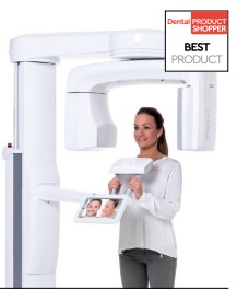
The Planmeca Viso G7 is designed to surpass the demands of industry leaders, specialists, and institutions. It has 4 built-in cameras and can capture a 30 × 30 cm volume and image from the skullcap to the cervical spine. The unit is ready to handle Planmeca ProFace and Planmeca 4D Jaw Motion technology, while the occipital head support allows an unimpeded view of facial tissue. It includes Planmeca CALM and Planmeca Ultra Low Dose technology. Patient positioning is done directly from the Planmeca Viso’s control panel using integrated cameras and a live patient view. Users can see the patient live from the control panel for flexible and exact FOV positioning. The volume size can also be adjusted freely

Dr. Clark has a general dental practice in downtown Nashville with a special interest in esthetics, TMJD, and sleep medicine. By completing over 1,500 hours of post-doctorate education, he excels at providing his patients with conservative therapies to improve esthetics, reduce pain and disease, and improve their overall health and well-being. Dr. Clark’s unique background and education, as well as his investment in cutting-edge technology, allow him to more effectively diagnose, treat, and manage the bite and the entire head and neck system for optimal patient outcomes. He earned his DMD from the University of Mississippi Medical Center and is an Associate Fellow at the World Clinical Laser Institute and a Fellow at the Las Vegas Institute for Advanced Dental Studies.
